Category Archives: Alzheimer Disease

High Intensity Exercise May Be Uniquely Beneficial to the Brain

…at least according to Dr Rhonda Patrick. I’d never heard of her before and don’t know her credentials.

You know what else is good for the brain? The Mediterranean diet.

Not Dr Patrick

I’m linking this video here in hopes of digging deeper in the future. You can read about her at what I assume is one of her websites.

Steve Parker, M.D.

Low to Moderate Alcohol Drinking Linked to Improved Cognitive Function in Adults

Jameson’s in a hotel bar near Chicago

From JAMA network:

These findings suggested that low to moderate alcohol drinking was associated with better global cognition scores, and these associations appeared stronger for white participants than for black participants.

Source: Association of Low to Moderate Alcohol Drinking With Cognitive Functions From Middle to Older Age Among US Adults | Dementia and Cognitive Impairment | JAMA Network Open | JAMA Network

     Steve Parker, M.D.

front cover of paleobetic diet

Click to purchase at Amazon.com. E-book also available at Smashwords. com.

Avoid Soybean Oil (if you’re a male mouse)

Soybean oil seems to be a real problem for male mice. We need more research in humans before declaring it a dangerous toxin to us. If you’re eating the Standard American Diet, you’ll find it hard if not impossible to avoid.

From EurekAlert:

New UC Riverside research shows soybean oil not only leads to obesity and diabetes, but could also affect neurological conditions like autism, Alzheimer’s disease, anxiety, and depression.

Used for fast food frying, added to packaged foods, and fed to livestock, soybean oil is by far the most widely produced and consumed edible oil in the U.S., according to the U.S. Department of Agriculture. In all likelihood, it is not healthy for humans.

Source: America’s most widely consumed oil causes genetic changes in the brain | EurekAlert! Science News

Steve Parker, M.D.

Steve Parker MD, Advanced Mediterranean Diet

Click the pic to purchase at Amazon.com. E-book versions also available at Smashwords. com.

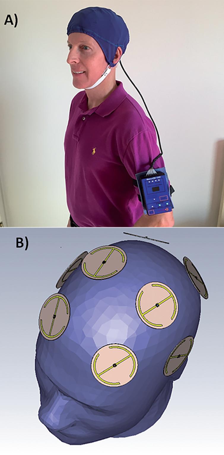
Transcranial Electromagnetic Treatment in Alzheimer’s Disease: Cognitive Enhancement and Associated Changes in Cerebrospinal Fluid, Blood, and Brain Imaging 

A) A MemorEMTM head device being worn by a subject. B) Position of the eight electromagnetic emitters embedded between the device’s two-layered head cap. Emitters collectively provide global forebrain TEMT via rapid sequential emitter activation.

This is from the cited journal article. I hope this is considered fair use rather than copyright infringement.

This is the most creative therapeutic approach to Alzheimer’s Disease I’ve seen in a while. It may even be preventative. I have no idea whether it will pan out in the long run. I’m always skeptical.

Click for a Scientific American article that discusses a different experimental protocol and probably different device.

Abstract

Background: Small aggregates (oligomers) of the toxic proteins amyloid-β (Aβ) and phospho-tau (p-tau) are essential contributors to Alzheimer’s disease (AD). In mouse models for AD or human AD brain extracts, Transcranial Electromagnetic Treatment (TEMT) disaggregates both Aβ and p-tau oligomers, and induces brain mitochondrial enhancement. These apparent “disease-modifying” actions of TEMT both prevent and reverse memory impairment in AD transgenic mice.

Objective: To evaluate the safety and initial clinical efficacy of TEMT against AD, a comprehensive open-label clinical trial was performed.

Methods: Eight mild/moderate AD patients were treated with TEMT in-home by their caregivers for 2 months utilizing a unique head device. TEMT was given for two 1-hour periods each day, with subjects primarily evaluated at baseline, end-of-treatment, and 2 weeks following treatment completion.

Results: No deleterious behavioral effects, discomfort, or physiologic changes resulted from 2 months of TEMT, as well as no evidence of tumor or microhemorrhage induction. TEMT induced clinically important and statistically significant improvements in ADAS-cog, as well as in the Rey AVLT. TEMT also produced increases in cerebrospinal fluid (CSF) levels of soluble Aβ1-40 and Aβ1-42, cognition-related changes in CSF oligomeric Aβ, a decreased CSF p-tau/Aβ1-42 ratio, and reduced levels of oligomeric Aβ in plasma. Pre- versus post-treatment FDG-PET brain scans revealed stable cerebral glucose utilization, with several subjects exhibiting enhanced glucose utilization. Evaluation of diffusion tensor imaging (fractional anisotropy) scans in individual subjects provided support for TEMT-induced increases in functional connectivity within the cognitively-important cingulate cortex/cingulum.

Conclusion: TEMT administration to AD subjects appears to be safe, while providing cognitive enhancement, changes to CSF/blood AD markers, and evidence of stable/enhanced brain connectivity.

Source: A Clinical Trial of Transcranial Electromagnetic Treatment in Alzheimer’s Disease: Cognitive Enhancement and Associated Changes in Cerebrospinal Fluid, Blood, and Brain Imaging – IOS Press

Steve Parker, M.D.

PS: I notice that several of the study authors are based in my area of operations, south-central Arizona.

PPS: The Mediterranean diet for years has been linked to lower risk of dementia.

Steve Parker MD, Advanced Mediterranean Diet

Click the pic to purchase at Amazon.com

If Gut Bacteria Cause Alzheimer’s Dementia, What If You Alter Those Bacteria?

The short answer? We don’t know the answer to either of those questions.

Low-carb salad

The gut bacteria (aka microbiome) seem to be able to decrease or increase inflammation that could cause or exacerbate Alzheimer’s dementia. The  microbiome’s effect on inflammation depends on the species of bacteria present, and the amount of those bacteria. At least one study found that Alzheimer’s patients have a greater abundance of the pro-inflammatory species and less of the anti-inflammatory species, compared to other folks.

Researchers with Wake Forest School of Medicine tried to find answers to the questions in the title of this post. (Click for full text.) They studied 17 experimental subjects, average age 64, who had mild cognitive impairment (11) or “cogni/subjective memory complaints” (6). God bless them for submitting to three spinal taps apiece. The experimental diets were 1) Mediterranean-Ketogenic (under 20 g carb/day), or 2) Low-fat American Heart Association diet (under 40 g fat/day). Participants were on each diet for six weeks.

The investigators didn’t find anything useful for those of us trying today to avoid Alzheimer’s or prevent the progression of mild cognitive impairment to dementia. Their bottom line is, “The data suggest that specific gut microbial signatures may depict [characterize] the mild cognitive impairment and that the modified Mediterranean-ketogenic diet can modulate the gut microbiome and metabolites in association with improved Alzheimer’s disease biomarkers in cerebrospinal fluid.”

So we won’t know for several more years, if ever, whether intentional modification of diet will “improve” our gut microbiomes, leading to lower risk of dementia.

What we have known for many year, however, is that the traditional Mediterranean diet is linked to lower risk of Alzhiemer’s dementia.

For more details, see Science Daily:

In a small pilot study, the researchers identified several distinct gut microbiome signatures — the chemicals produced by bacteria — in study participants with mild cognitive impairment (MCI) but not in their counterparts with normal cognition, and found that these bacterial signatures correlated with higher levels of markers of Alzheimer’s disease in the cerebrospinal fluid of the participants with MCI.

Through cross-group dietary intervention, the study also showed that a modified Mediterranean-ketogenic diet produced changes in the gut microbiome and its metabolites that correlated with reduced levels of Alzheimer’s markers in the members of both study groups.

Source: Diet’s effect on gut bacteria could play role in reducing Alzheimer’s risk — ScienceDaily

Steve Parker, M.D.

Steve Parker MD, Advanced Mediterranean Diet

Two diets in one book, including the Ketogenic Mediterranean Diet

Click the pic to purchase the world’s first practical ketogenic Mediterranean diet at Amazon.com


Can a Ketogenic Diet Prevent or Treat  Alzheimer’s disease?

Sunny’s Super Salad

Maybe…we don’t know yet. From a recent scientific article:

“Highlights

•Impaired brain glucose metabolism and amyloid β plaques are associated with Alzheimer’s disease pathology.

•Ketones provide an alternative metabolic precursor to glucose in the brain.

•Ketogenic diets likely reduce amyloid plaques and may reverse their neurotoxicity.

•Modern diets high in carbohydrates may contribute to increasing Alzheimer’s incidence.

•The ketogenic diet (including carbohydrate restriction) might be useful in the management of Alzheimer’s disease.”

Source: The ketogenic diet as a potential treatment and prevention strategy for Alzheimer’s disease – ScienceDirect

Poor Sleep Linked to Alzhieimer’s Disease

“A 2017 analysis combined results of 27 studies that looked at the relationship between sleep and cognitive problems, including Alzheimer’s. Overall, poor sleepers appeared to have about a 68 percent higher risk of these disorders than those who were rested, researchers reported last year in Sleep. That said, most studies have a chicken-and-egg problem. Alzheimer’s is known to cause difficulty sleeping. If Alzheimer’s both affects sleep and is affected by it, which comes first?For now, the direction and the strength of the cause-and-effect arrow remain unclear. But approximately one-third of U.S. adults are considered sleep deprived (getting less than seven hours of sleep a night) and Alzheimer’s is expected to strike almost 14 million U.S. adults by 2050 (5.7 million have the disease today). The research has the potential to make a big difference.”

Source: The brain may clean out Alzheimer’s plaques during sleep | Science News

Yes, Seafood Consumption Will Contaminate Your Brain With Mercury, But It Doesn’t Seem to Cause Damage 

Dead whole fish aren't very appealing to many folks

Dead whole fish aren’t very appealing to many folks

I advocate consumption of cold-water fatty fish a couple times per week for long-term protection against heart and brain disease. The protective component of fish may be the omega-3 fatty acids.

On the other hand, much seafood is contaminated with mercury, which can be toxic. So, is the mercury in fish actually toxic to brain tissue of folks eating reasonable amounts of fish?

A recent autopsy study answers, “No.”

Read further for details.

Much more appetizing!

From the Journal of the American Medical Association, 2016 Feb 2;315(5):489-97. doi: 10.1001/jama.2015.19451. “Association of Seafood Consumption, Brain Mercury Level, and APOE ε4 Status With Brain Neuropathology in Older Adults.”

IMPORTANCE:Seafood consumption is promoted for its many health benefits even though its contamination by mercury, a known neurotoxin, is a growing concern.

OBJECTIVE:To determine whether seafood consumption is correlated with increased brain mercury levels and also whether seafood consumption or brain mercury levels are correlated with brain neuropathologies.

DESIGN, SETTING, AND PARTICIPANTS:Cross-sectional analyses of deceased participants in the Memory and Aging Project clinical neuropathological cohort study, 2004-2013. Participants resided in Chicago retirement communities and subsidized housing. The study included 286 autopsied brains of 554 deceased participants (51.6%). The mean (SD) age at death was 89.9 (6.1) years, 67% (193) were women, and the mean (SD) educational attainment was 14.6 (2.7) years.

EXPOSURES:Seafood intake was first measured by a food frequency questionnaire at a mean of 4.5 years before death.

MAIN OUTCOMES AND MEASURES:Dementia-related pathologies assessed were Alzheimer disease, Lewy bodies, and the number of macroinfarcts and microinfarcts. Dietary consumption of seafood and n-3 fatty acids was annually assessed by a food frequency questionnaire in the years before death. Tissue concentrations of mercury and selenium were measured using instrumental neutron activation analyses.RESULTS:Among the 286 autopsied brains of 544 participants, brain mercury levels were positively correlated with the number of seafood meals consumed per week (ρ = 0.16; P = .02). In models adjusted for age, sex, education, and total energy intake, seafood consumption (≥ 1 meal[s]/week) was significantly correlated with less Alzheimer disease pathology including lower density of neuritic plaques (β = -0.69 score units [95% CI, -1.34 to -0.04]), less severe and widespread neurofibrillary tangles (β = -0.77 score units [95% CI, -1.52 to -0.02]), and lower neuropathologically defined Alzheimer disease (β = -0.53 score units [95% CI, -0.96 to -0.10]) but only among apolipoprotein E (APOE ε4) carriers. Higher intake levels of α-linolenic acid (18:3 n-3) were correlated with lower odds of cerebral macroinfarctions (odds ratio for tertiles 3 vs 1, 0.51 [95% CI, 0.27 to 0.94]). Fish oil supplementation had no statistically significant correlation with any neuropathologic marker. Higher brain concentrations of mercury were not significantly correlated with increased levels of brain neuropathology.

CONCLUSIONS AND RELEVANCE:In cross-sectional analyses, moderate seafood consumption was correlated with lesser Alzheimer disease neuropathology. Although seafood consumption was also correlated with higher brain levels of mercury, these levels were not correlated with brain neuropathology.

Source: Association of Seafood Consumption, Brain Mercury Level, and APOE ε4 Status With Brain Neuropathology in Older Adults. – PubMed – NCBI

Lithium as a Preventative or Treatment of Alzheimer’s Disease

The following excerpt is boring, so move along now. It’s from a recent review article on all the potential causes and therapeutic options for Alzheimer’s Disease (AD). I post it here so I can find it later. Click the link at bottom to RTWT.

“Lithium is not yet generally recognized as a trace element but several lines of evidence make it a strong candidate. For instance, long-term low-dose exposure to lithium exerts anti-aging capabilities and unambiguously decreases mortality in animal models. In humans, epidemiological studies indicate an inverse correlation between lithium concentration in drinking water and mood, depression and suicide rates, amongst other psychiatric conditions. In a study that compared elderly bipolar patients (who exhibit a higher risk for dementia) who had received chronic lithium treatment, with bipolar patients who had not received lithium, it was shown that the prevalence of the treated group was equivalent to the general, age-comparable population, whereas the non-lithium-treated patients had an incidence of dementia that was six times greater, i.e. 5 % vs. 33 %, respectively. In another study it was shown that lithium treatment resulted in an increase in volumes of the hippocampi in both hemispheres compared to an unmedicated group, an effect that was apparent even after a brief treatment period of about 4 weeks on average. Importantly, intake of lithium not only in standard therapeutic but also in trace doses reduces the risk for dementia, suicide, and other behavioural outcomes, suggesting an pharmacological interference with key regulators of these pathological processes. So, lithium naturally regulates critical cell signalling pathways and a lack of lithium in the diet can therefore cause increased disease risk.It has been shown that lithium modulates negatively the activity of the two kinases GSK-3α and GSK-3β, which might explain both the relative specificity and sensitivity of the effects of low-dose lithium treatment (see below). Since GSK-3β-activation by oligomeric Aβ promotes neuroinflammation, phosphorylation of tau and disturbance of AHN [adult hippocampal neurogenesis], all key mechanisms in the AD process, in inhibition of GSK-3 by lithium results in reduced tauopathy and neurodegeneration in vivo. Likewise, lithium treatment was shown to improve AHN, neuropathology and cognitive functions in a mouse model of AD. Furthermore, such “AD-mice” treated from two months of age had decreased numbers of senile plaques, no neuronal loss in cortex and hippocampus and increased BDNF levels when compared to non-treated transgenic mice. In order to achieve this effect, it was sufficient to give lithium at about one per mill of the high standard-dose therapy in bipolar disorder, a dose which can cause some significant side effects. Hence the authors of the study believe that their data support the use of (virtually side-effect free) microdose lithium in the prevention and treatment of Alzheimer’s disease.Indeed, long-term lithium treatment already provided preliminary evidence of its disease-modifying properties for amnestic MCI [mild cognitive impairment] in a randomised controlled trial, where the lithium-treated group had fewer conversions from MCI to AD. Lithium treatment was associated with a significant decrease in cerebrospinal fluid concentrations of hyperphosphorylated tau and better performance on the cognitive subscale of the Alzheimer’s Disease Assessment Scale and in attention tasks. At a more advanced stage of AD, microdose treatment with only 300 μg lithium administered once daily stabilized the AD patients during the complete evaluation phase of 15 months. For instance, whereas the treated group showed no decreased performance in the mini-mental state examination (MMSE), lower scores were observed for the control group during the same period, with significant differences occurring after three months, and increasing progressively.Importantly, lithium, besides being required for efficient AHN and blocking AD-specific pathological processes, also impacts on cell-rejuvenating autophagy. Lithium was found to inhibit the activity of inositol monophosphatase (IMPase), which leads to a decrease of myo-1, 4, 5-triphosphate (IP3). This reduction of IP3-activity induces autophagy, independent of mTOR. In this context it is important to note that lithium chloride extends the lifespan of the nematode Caenorhabditis elegans, possibly by means of mitochondrial rejuvenation, which suggests that lithium exerts its effects on evolutionary highly conserved mechanisms. Intake of drinking water with comparable low lithium concentrations were found to be inversely correlated with all-cause mortality in a large epidemiological study in Japan. Hence a lack of lithium is linking aging and frailty (all increasing mortality) to disturbed autophagy, and AD to impaired AHN, and, thus, might represent another important and modifiable risk factor in this neurodegenerative disease. Traces of lithium can be ingested in some geographic areas by drinking local tap water or otherwise by consuming commercially available, mineral-rich spring waters, containing suitable concentrations of around 1 mg lithium per litre. Hence microdose lithium intake by means of one or two glasses of such water a day is not only of potential therapeutic value (see below) but also a safe preventive measure, by means of simply reducing an intake-deficit of an important novel trace element.”

Source: Unified theory of Alzheimer’s disease (UTAD): implications for prevention and curative therapy

Bonus excerpt:

“Taken together, besides the importance of IMF and physical exercise, also dietary MCTGs [medium-chain trigylcerides] can be an attractive (indirect) source of ketone bodies during the non-fasting state. The intake of coconut oil as a healthy source of MCTGs has additional positive effects on neuronal insulin resistance, neuroinflammation as well as Aβ-toxicity, improves the LDL/HDL-cholesterol quotient by increasing HDL, and ameliorates several others key progression factors of AD. Hence the use of virgin coconut oil is highly recommended as a safe and healthy alternative for polyunsaturated oils for frying and baking and butter and an important part of a comprehensive strategy for the prevention and treatment of AD.”

Unified theory of Alzheimer’s disease (UTAD): implications for prevention and curative therapy

No need to read the following. It’ll likely bore you to death. I record it here for my own purposes. Alzheimer’s Disease is a huge problem and we desperately need ways to prevent and cure it. Prevention should be easier than cure. BTW, the Mediterranean diet is linked to lower risk of Alzheimer’s Disease.

“The master regulator/inhibitor of autophagy is the mammalian target of rapamycin (mTOR). This intracellular kinase functions as a key signalling node that integrates information regarding extracellular growth factor stimulation, nutrient availability and energy supplies. The fungal metabolite rapamycin was accidently found to block mTOR and became not only the eponym of mTOR but also the main molecular tool to dissect mTOR-function. Rapamycin treatment was found to activate autophagy by inhibiting mTOR, thereby slowing down both aging and cognitive decline of caged mice, suggesting that inefficient autophagy as part of the NRJ-program might be a central element of both processes. Conversely, caging (standard housing) might eliminate an important behavioural cues (like for instance physical activity or intermittent fasting, see below), which leads to unnaturally high activity of mTOR and low activity of peroxisome proliferator-activated receptor γ coactivator 1α (PGC-1α), the master controller of mitochondrogenesis (see below), thereby inhibiting neuron-rejuvenating and protecting autophagy. Hence the observed aging and cognitive decline in murine models of AD might be regarded as artificial and not reflecting the aging process under natural conditions.

Other behavioural cues that inactivate mTOR are, besides physical exercise, which was shown to promote autophagy of defunct organelles and macromolecules in the brain, chronic caloric restriction (CCR), which is well known to delay aging and extend life-span in essentially all eukaryotic organism. But is CCR a physiological cue or rather an artefact of experimental research that simulates, to some extent, a more natural dietary pattern, namely intermittent fasting (IMF), which from an evolutionary point of view is more natural (see below)? But IMF is experimentally more labour-intensive than CCR and therefore less well studied. Nevertheless, autophagy and in particular mitophagy was found to being activated by CCR through inhibition of mTOR in essentially all species investigated, ranging from yeast, to flies, worms, fish, rodents and even to rhesus monkeys [87], thereby decelerating mTOR-driven aging. CCR not only extends lifespan, it also protects the central nervous system from neurodegenerative disorders, whereas excessive caloric intake is clearly associated with accelerated aging of the brain and increased the risk of neurodegenerative disorders due to suppressed autophagy.

Nevertheless, IMF was shown to create a more robust and steady inhibition of mTOR-accelerated aging and cognitive decline when compared to CCR. This is explained by the fact that the main hormone-like signalling molecules of the metabolic status during IMF, the ketone bodies acetoacetate (AcAc) and D-β-hydroxybutyrate (βOHB), are more efficiently generated during fasting than by CCR. These two respiratory fuels can endogenously be produced by the liver in large quantities (up to 150 g/day) from mobilized fatty acids in a variety of physiological or pathological conditions. In humans, basal serum levels of βOHB are in the low micromolar range, but rise up to several hundred micromole after 12 to 16 h of fasting. Importantly, when blood glucose and insulin are low, up to 60 % of the brain energy needs can be derived from ketone bodies, replacing glucose as its primary fuel. Similar high levels of up to 1 to 2 millimole βOHB are reached after prolonged endurance exercise. A physiologically relevant increase in ketone body production is already achieved by fasting overnight, which can even be enhanced if we are physically active before breaking the fasting in the morning. This most likely mimics the situation that faced our foraging ancestors who went out for hunting or gathering food with their stomachs empty.

Since neither long- nor medium-chain saturated fatty acids can pass the BBB, only their transformation into ketone bodies allows our energy-demanding brain to access the largest energy store, our adipose tissue. In fact, ketone body production reduces glucose requirement and preserves gluconeogenic protein stores during fasting, which enables a profound increase in the capacity for survival. Interestingly and again in line with the GMH, elderly generate ketone bodies at least as efficient as younger adults during IMF and the metabolic response to a ketogenic diet appears also to be unaffected by aging.As hinted at above, the observation that IMF is superior to CCR makes also a lot of sense from an evolutionary perspective, as not chronic starvation but rather periodic alteration between fasting and intake of high-caloric meals after successful foraging was ancient normality. Importantly, recent evidence suggests that our phylogenetically conserved genetic program uses the metabolic changes that originate from intermittent fasting (IMF) as a behavioural cue of for [sic] the initiation of subcellular renewal. This is a good thing, since in order to maintain cellular youth, we do not have to starve by CCR. It is sufficient to alternate phases of fasting, which just need to be sufficiently long to induce ketone body production (for instance 12 h overnight) and phases of eating, in which the total energy demand of our body can be met. In contrast, current normality consists of constant feeding pattern, which results in permanent high mTOR activity (and low PGC-1α-levels, see below), which suppresses cellular rejuvenation. A sedentary lifestyle aggravates this pro-aging effect, whereas prolonged physical exercise reduces mTOR-activity, possibly also by increasing ketone body production.”

Source: Unified theory of Alzheimer’s disease (UTAD): implications for prevention and curative therapy